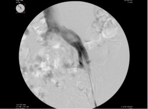
Bi-planar contrast venography from a direct venous puncture is a common investigation in patients with suspected non-thrombotic iliac venous lesions (NIVLs). Some authors have proposed that intravascular ultrasound (IVUS), also via a direct venous puncture, should be used as the gold standard to identify and characterize iliac vein lesions prior to stenting. Recently, the requirement for IVUS has been challenged because it may not provide further useful information in the majority of patients. It may be possible to identify a sub-group who can proceed direct to stenting from venographic appearances.
ANIL HINGORANI & ENRICO ASCHER When assessing the supra-inguinal veins in patients with chronic venous disease, it is generally agreed that IVUS is mandatory. In an attempt to identify a sub-group of patients where the use of IVUS could be averted, we analyzed and evaluated the images of patients who had both standard contrast venograms and IVUS exams. We compared venographic images and IVUS scans in 92 legs. The appearances of the veins were classified as follows:
Type I: Normal to mild vein narrowing or dilatation ≤ 20% compared to the adjacent segment.
Type II: Moderate ≥ 21% and ≤ 40%.
Type III: Severe ≥ 41%.
Type IV: Bull’s eye sign.
The Bull’s eye sign is a radiological appearance defined as a central circle with minimal or no dye within a dilated vein and forking of the dye around the circle. This is in concordance with the congenital adhesions in the common iliac vein described by the anatomist J. Playfair McMurrich in 1906 on cadavers. Of the 92 venograms we studied, 88 had a > 50 % diameter or cross sectional area reduction on IVUS. The correlation of venographic findings and a positive IVUS scan was as follows:
Type I (26) had 85% positive IVUS; Type II (22) had 100% positive IVUS; Type III (25) had 100% positive IVUS and Type IV (19) had 100% positive IVUS. This suggests that IVUS was useful only in identifying positive lesions in iliac veins that had ≤ 20% vein narrowing on venography.
The new proposed classification of venographic findings can be used to treat more than two thirds of the patients with NIVLs without resorting to the use of IVUS.
Venography image demonstrating the Bull’s eye sign (Type IV). A central circle is shown with minimal or no contrast within the dilated left common iliac vein. Contrast is seen forking around this circle.
