The clinical part of the CEAP (Clinical, Etiological, Anatomical, Pathological) classification is the most popular classification system for venous disorders. Its purpose originally in 1994 was to standardise reporting by defining the study groups in clinical trials and manuscripts submitted to journals. However, the C of CEAP is not a perfect system and there have been several recommendations for change. There are 3 possible directions available to improve clinical CEAP: to extend, to revise or to replace. The French group propose an extension to the existing classification which is backward compatible and more informative.
ANDRÉ CORNU-THENARD Many phlebologists and other specialties use the CEAP classification in order to classify their venous patients into groups. In France this was necessary because of the numerous studies performed on venous populations, epidemiological research, symptoms and signs as well as many studies on the CEAP classification itself. During this work it became apparent that there are several unresolved problems in using the clinical part of the CEAP classification. Two examples are provided below.
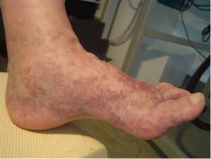
(i) Corona Phlebectatica Paraplantaris (CPh, Fig 1) is the appearance of blue/red venules and telangectasiae of the foot. These would be classified as C1 under the existing system. However, it has been shown that the presence of CPh is a more advanced stage of chronic venous disorder and that it is predictive of the development of venous ulceration.
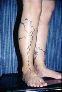
(ii) All kinds of varicose veins (VVs) are included in C2. The clinical features, pathology and evolution of recurrent VVs are very different from primary VVs which should be reflected in a new system. Furthermore, recurrent VVs are the focus of the REVAS (Recurrent VVs After Surgery) and PREVAIT (VVs after any potential curative treatment, residual/recurrent or both) (Fig2).
In collaboration with Patrick H Carpentier and Jean-François Uhl we decided that a change was necessary and proposed an extension to the existing classification. This was performed without any change to the basic structure with a similar organizational layout at all the respective levels. The suggestion was to provide more information by subdividing each of the existing ‘C’ parts into Ca and Cb. This follows on from the first revision in 2001 when C4 was subdivided into C4a (Pigmentation) and C4b (Lipodermatosclerosis, LDS).
C0a = Asymptomatic C0s = Symptomatic
C1a = Telangectasiae C1b = Reticular blue veins
C2a = Primary VVs C2b = Recurrent VVs
C3a = Oedema C3b = Corona Phlebectatica
C4a = Eczema, Pigmentation C4b = LDS, Atrophie Blanche
C5a = Venous ulcer healed C5b = Recurrent venous ulcer healed
C6a = Venous ulcer C6b = Recurrent venous ulcer
Fig 1, Corona Phlebectatica. The most part of these telangectasiae are situated on the lateral sides of the foot but when they became larger they can be found on the planter and dorsal sides.
Fig 2, duplex mapping of recurrent VVs after GSV stripping with VVs 5 years later. The 3 small circles signify perforating veins.

