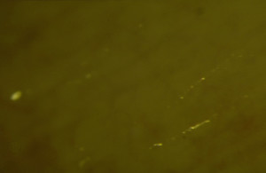
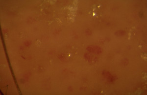
This is the examination of the microcirculation using a microscope at a magnification in the order of 50 X. It has the potential to detect the cutaneous manifestations of chronic venous insufficiency (CVI) before overt skin changes are seen clinically. The technique has been described as a microscopic C of CEAP in the assessment of disease severity. The underlying hypothesis is that venous micro-angiopathy is the central pathophysiology in the development of cutaneous complications. The general use of capillaroscopy may complement the clinical and duplex assessments of venous disease thereby providing a more complete picture of severity.
ROMEO COSTANZO MARTINI Capillaroscopy is a noninvasive investigation test for the assessment of the skin microcirculation. It is routinely performed utilizing a video camera with an image magnification between 50 and 100 X. Capillaroscopy can be performed along the entire skin of the body surface but it is usually performed at the nailfold of the finger (or toe?). Here the capillaries loops run parallel to the skin surface and can be seen easily. However, on other parts of the body, only the apex part of the capillary tufts may be observed.
The morphological analysis of capillaries includes the assessment of their diameter, density and architectural arrangement, as well as the peri-capillary tissue and the way in which the blood flows inside the capillaries. In the more advanced stages of CVI typical glomerular capillaries are visible. The glomerular capillaries may be assumed as an early damage of the skin microcirculation induced by venous macrovascular hypertension.
Early stage of CVI in a woman with varicose veins without skin changes.
Advanced CVI with venous eczema and fibrosis. Note the presence of glomerular like capillaries.

