


Anatomical dissections provide a unique insight into how veins function. Whilst they cannot demonstrate flow patterns resulting from muscular contraction they are able to illustrate the pathways available for drainage. Flow direction in health can be inferred by the direction of the valves.
The foot venous pump (FVP) is a significant contributor to augmenting the venous return. Haemodynamic studies suggest that 25% of the venous return is provided by the FVP and the rest by the calf muscle pump (CMP). A failing FVP has been attributed to foot arch abnormalities and ankle ankylosis which may be significant contributors to the development of venous skin changes. Furthermore, venous ulcers appearing below their usual location in the gaiter region, inferior to the malleoli, suggest failure of the FVP. The haemodynamics of the FVP is a grossly under researched area.
CLAUDE GILLOT The perforating veins (PVs) of the foot are shown in red (7, 8, 9 & 10) and represent the outflow of the foot pump into both saphenous systems. Courtesy of J-F UHL.
The heart of the FVP is located deep in the foot in the plantar veins (5 & 6). Like the soleal venous sinuses, these veins appear capacitance vessels. Their outflow goes into the posterior tibial veins (4) and anterior tibial veins (not shown). However, the most interesting observation is the presence of outflow connections from the foot PVs into both saphenous systems. The valves within these veins suggest that the normal direction of flow is outward from deep to superficial. This is in direct contrast to the direction of the valves in the calf PVs, which are often paired, and suggest that the normal flow direction is inward, from superficial to deep.
The great saphenous vein (1) originates below the medial malleolus from 3 roots: the medial marginal vein (3), the sub-malleolar PV (7) and the dorsal PV of the foot (not shown). The PV of the first metatarsal space gives rise to the medial marginal vein which receives 2 medial PVs: the navicular (8) and cuneiform PV (9). These veins constitute the medial drainage pathways of the foot.
The small saphenous vein (2) is fed by the inter-tendinous PV and an Achilles tributary coming from the PV of the calcaneus (10) which is a direct outflow of the lateral plantar veins. These constitute the lateral drainage pathways of the foot.
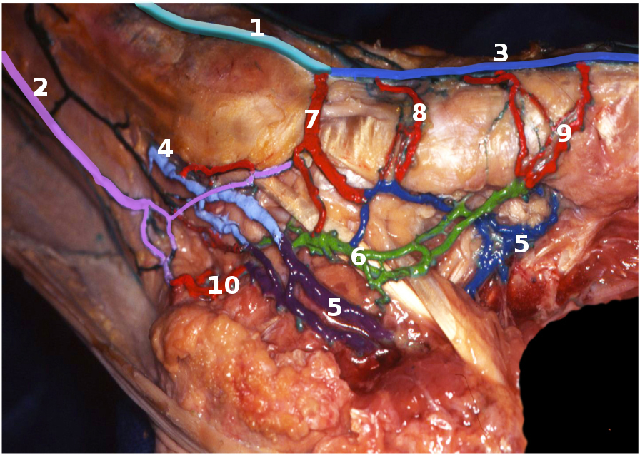
Anatomy of the foot venous pump (FVP) illustrating the drainage pathways. The medial structures have been reflected back to expose the small saphenous vein (2).